VisiTech iSIM
Manufacturer:
VisiTech
Model name:
VisiTech iSIM
Location:
HPM D34.1
Phone:
1 23 45
Instrument contact:
ScopeM
Otto-Stern-Weg 3
8093
Zürich
Switzerland
Brief description
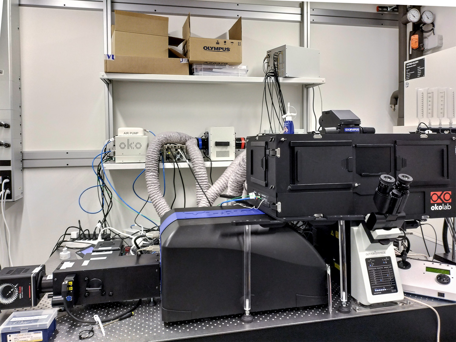
iSIM (or instant Scanning Image Microscopy) system from the company external page VisiTech is an analog implementation of Image Scanning Microscopy (as will be described below), generating a super-resolution image (X, Y resolution of ~120 nm and Z resolution of ~280 nm ) in real-time, with no requirements for post-processing. It is mostly suited for high-speed, live-cell imaging applications.
Based on the so-called pixel re-assignment method, super-resolution imaging is performed by a single optical element in the emission path (μLens array), which re-assigns the image information in real-time.
The iSIM technology is integrated into a state-of-the-art Evident (formerly Olympus) microscope platform. It combines ultra-fast 3D imaging with low phototoxicity and provides close to a two-fold resolution enhancement in X, Y and Z, compared to standard confocal or wide-field fluorescence microscopy.
Applications
- Fast and gentle live super-resolution (X, Y resolution of ~120 nm and Z resolution of ~280 nm) imaging in 2D and 3D
- Two colour simultaneous live super-resolution imaging
- Multi-channel super-resolution on fixed samples
Technical details
Microscope Frame:
- Evident IXplore IX83 inverted microscope with Temperature Control and gas control
Available objectives:
- 100x/NA 1.5 UPLAPO100XOHR Olympus objective
- 20x/NA 0.75 UPlanSApo Olympus objective
Cameras:
- 2x Hamamatsu ORCA-QUEST (QE around 95 %) with USB interface
Available laser lines:
- 405 nm
- 488 nm
- 561 nm
- 640 nm
- 730 nm
Widefield illumination:
- pE 300 white light LED
Microscope stage:
- Piezo scanning ASI stage with insets for slides (and IBIDI chambered coverslip slide formats), Labtek chambered coverglass formats, 35 mm dishes and multiwell plates
Software:
- CellSens Dimension
Specifications:
- Large FOV Up to ~13.3 mm × 13.3 mm (176.9 mm2)
- Spatial & axial resolution achieved across Large FOV ~120 nm later resolution, and ~280 nm axial resolution
- Temporal Resolution Capable of 100 fps (camera dependant)
- Broadband Uniform and Homogeneous Illumination across Large FOV Corner to Corner Roll Off: <6 % 488–640 nm, <20 % 405 nm
- Automated bright-field by-pass available
- No requirement for intermediary magnification results in super resolution images being generated without any loss in signal and with very low levels of photo-bleaching
- Re-assignment micro-lens array is positioned after pinhole array in emission path so as to ensure optimal confocality
- Emission pin hole array is in first conjugate plane to reduce imaging cross-talk and spherical aberrations
Extra features:
- Simultaneous multi-colour imaging of up to 4 fluorescent colours, each with a field of view of 130 μm × 130 μm @100x with DUAL Camera option
- Combination with incubators for live-cell imaging and FRAP under the control of cellSens software
Further documents:
Further information about the instrument can be found in these documents: