Olympus/Evident VS200 Slide Scanner
Manufacturer:
Olympus/Evident
Model name:
Olympus/Evident SlideviewTM VS200 Slide Scanner
Location:
HPL E35.2
Phone:
3 33 42
Instrument contact:
Dr. Gabriella Éva Bodizs
ScopeM
Otto-Stern-Weg 3
8093
Zürich
Switzerland
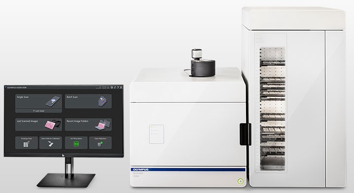
Brief description
The Evident/Olympus VS200 slide scanner is an automated microscope for fast imaging of entire tissue sections designed to speed up and simplify the image acquisition of medium and large-scale histology projects.
Key features
- High resolution image of an entire tissue section (ex: mouse brain) in one file (due to the automated stitching) instead of multiple snapshots
- High scanning speed with established workflow settings for high-throughput imaging
- Up to 210 microscopy slides imaging in one run (35 trays exchanged by a robotic arm)
- Motorized colour and black&white cameras
- 5 flexible imaging modes: brightfield, polarization, fluorescence, darkfield, and phase contrast in one system, with the ability to combine multiple techniques in a single scan.
- Upright system with multiple air (2x, 4x, 10x, 20x, 40x) and oil objectives (40xOil) with integrated on-the-fly automatic oil dispenser
- Scanning possibilities in manual and/or automatic modes.
- Various simple-to-use slide tray supports: 26 × 76 mm, 52 × 76 mm, 76 × 102 mm, and 102 × 127 mm slide
Technical details
Microscope:
- Evident/Olympus SLIDEVIEV VS200
Imaging Methods:
- BF-brightfield, FL-fluorescence, POL-polarization, DF-darkfield, PH-phase contrast
Multi-band Filter sets:
- Penta-band filter set with DAPI/FITC/Cy3/Cy5/Cy7: external page DAPI/FITC/Cy3/Cy5/Cy7 Penta "Sedat" LED HC Set | F66-987
- Triple-band filter set with CFP/YFP/mCherry: external page CFP/YFP/mCherry Triple sbx LED HC set | F66-436
Objectives:
- 2x: PLN2x/ NA 0.06 / WD: 5,8 mm
- 4x: UPLFLN4X/ NA 0.13 / WD: 17 mm
- 10x: UPLXAPO10X/ NA 0.4 / WD: 3.1 mm
- 20x: UPLXAPO20X/ NA 0.8 / WD: 0.6 mm
- 40x: UPLXAPO40X/ NA 0.95 / WD: 0.18 mm
- 40x oil: UPLXAPO40XO/ NA 1.4 / WD: 0.13 mm
Camera/detectors:
- Colour camera: 5 Megapixel colour camera, Integrated 2/3-inch CMOS, 3.45 μm × 3.45 μm pixel size, high sensitivity, high resolution
- Black&White camera: Hamamatsu Orca-Fusion BT, sCMOS camera for C-mount adaptation, 2304 × 2304 pixels with 6.5 ×6.5 μm cell size
Light sources:
- LED light with built-in Köhler illumination for transmitted light, high-intensity and high colour rendering LED (up to 50,000 hours)
- X-Cite NOVEM 9-Channel LED Illumination: external page https://www.excelitas.com/product/x-cite-novem-powerful-9-channel-led-illumination-system
- Peaks: 385 nm, 435 nm, 475 nm, 500–600 nm, 530 nm, 555 nm, 580 nm, 635 nm, 735 nm
Scanning time:
- Approx. 80 sec (20X objective, scan area 15 mm × 15 mm (0.6 in. × 0.6 in.) brightfield)
Software:
- Slideview VS200-ASW – acquisition software
Automatic sample detection, automatic barcode reading, automatic focus mapping, automatic scanning, automatic stitching, pause and resume scanning, Z stack imaging extended focus imaging (EFI), multiple image formats (vsi, JPEG, and TIFF), synchronized multi-image display, stepless zooming, slide browsing while scanning, annotations, screen capture, slide loader control (multiple tray loader only) - OlyVia Free viewer software: external page https://evidentscientific.com/en/downloads