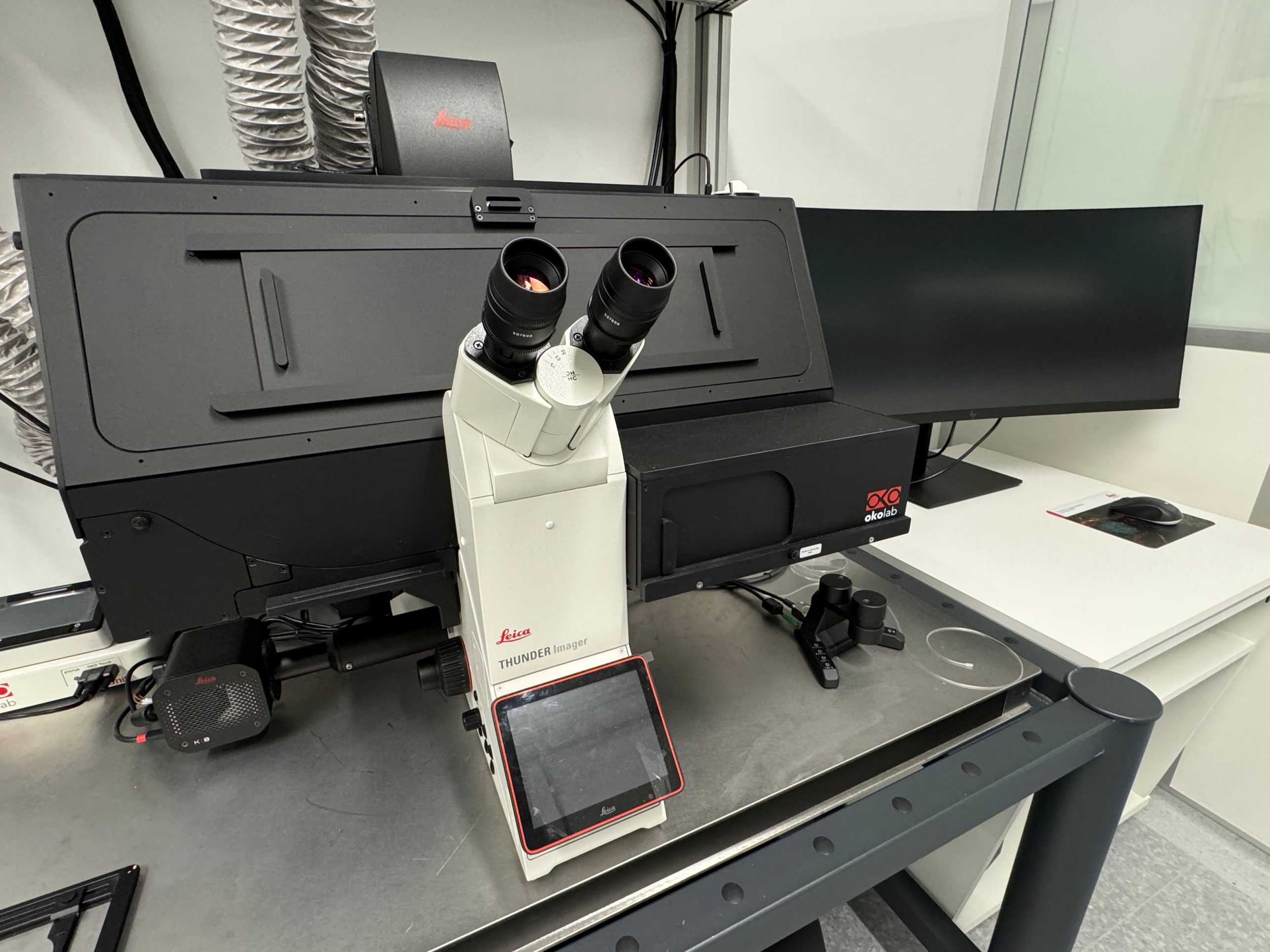
Leica Thunder
Manufacturer:
Leica
Model name:
Leica Thunder Imager Nano Cell
Location:
HCI H396.1
Instrument contact:
ScopeM
Otto-Stern-Weg 3
8093
Zürich
Switzerland
The performance advantage is achieved thanks to an opto-digital method developed by Leica called Computational Clearence that removes in real time the out of focus light usually present in widefield images allowing the system to collect 2D and 3D data with high contrast and high signal to noise ratio. The better SNR allows for thicker samples, about 150um, to be imaged and analysed. Furthermore, together with adaptive deconvolution there is a 2-fold enhancement of lateral resolution.
The Leica Thunder bridge the gap between traditional widefield and confocal microscopy providing the speed and sensitivity of a camera-based system together with an increased image quality allowing to for rapid time-lapse events to be captured with confocal-like contrast.
For live cell imaging, the microscope is also equipped with an incubator with temperature and CO2 control. Furthermore, the Thunder is also equipped with hardware-based adaptive focus control (AFC) for precise drift correction and Z adjustment during time-lapse applications.
Leica Thunder is also designed to be part of a light microscopy-electron microscopy correlative workflow designed by Leica called The Coral Life Workflow. By working in combination with the Leica’s EM-ICE High Pressure Freezer. The high-quality images obtained with the Thunder allow targeting your sample with regions-of-interest for downstream nano-level analysis in the electron microscope. The direct connection to the EM-ICE then allows time-point specific conservation of the sample for electron microscopy processing facilitating the investigation of dynamic events at nanometer resolution.
Technical details
Available objectives:
- HC PL FLUOTAR 20x/0.55 DRY
- HC PL APO 40x/1.1 WATER
Cameras:
- K8 CMOS Camera (QE around 95%)
Widefield illumination:
- Leica LED8
390 nm
440 nm
475 nm
510 nm
555 nm
575 nm
635 nm
747 nm
Filter settings:
Emission exteral filterwheel EFW_LED8:
- 460/80
- 535/70
- 590/50
- 642/80
- Transmission
Microscope stage:
- Quantum linear motor stage - high precision stage with insets for slides, petri dishes and multiwell plates.
Software:
- Leica LAX 3.9.1
