Leica TIRF
Brief description
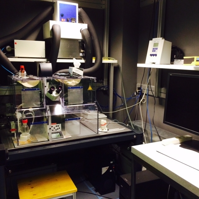
This epi-fluorescence microscope is additionally equipped with a TIRF unit enabling to view processes at the interface to the coverslip (adhesion, membrane fusion processes etc.). The system has a high speed function allowing for fast aquisition using the laser illumination. It can also be used for automated longterm, multi-position life cell imaging.
The setup includes a motorized X-Y stage, and an incubator box temperature and CO2). It is operated by Leica LAS AF software. An offline version of the program exists on Image Processing Computer 1 in G 9.1 for viewing images and further analysis (can also be downloaded onto your PC). The system is equipped also with an Eppendorf FemtoJet and InjectMan Ni2 microinjection unit, which allows semi-automatic microinjection of adherent cells.
Technical details
Microscope:
- Leica DMI6000B (inverted), equipped for DIC and Phase contrast imaging
Ilumination:
- Leica Metal-halide illumination source (similar spectra like an HBO) for conventional widefield epifluorescence
- laser lines: 405, 488, 561 and 647 nm for TIRF
Objectives:
- 5x 0.12NA N-Plan
- 10x 0.25NA Ph1 N-Plan
- 20x 0.4NA Ph1 HCX PlanFluotuar LD (long distance)
- 20x 0.7NA HC PlanApo
- 40x 0.7NA Ph2 PlanFluotar
- 63x 1.4NA Oil HCX PlanApo DIC
- TIRF objective: 100x 1,47NA Oil HCX PlanApo
Camera / detectors:
- Andor iXon EM CCD camera (front illuminated, 8 μm x 8 μm pixel size)
Software:
- Leica LAS AF software
Fluorescence settings:
- Widefield epifluorescence filtersets: DAPI , EGFP, Cy3, DsRed and Cy5