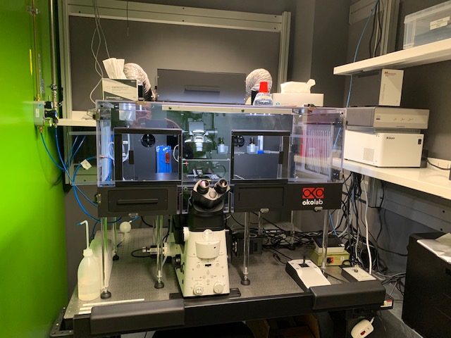
Nikon MMI Microdissection
Brief description
This microscope enables you to perform precise, non-contact laser based micromanipulation experiments. The MMI (Molecular Machines & Industries ) CellCut facilitates precise and contamination-free dissection of cell clusters, single cells or subcellular compartments from various types of tissues including fresh frozen, paraffin embedded and archived slides, cytospins, smears and even living cells. The system is operated by the MMI software.
Additionally, you are also able to do high resolution imaging of fixed stained and unstained samples and it is also possible to perform complex live cell imaging experiments. The "imaging functionality" is controlled by the Software NIS-Elements (Nikon) which allows multichannel, multiposition, time lapse imaging and complex experiments.
Technical details
Microscope:
- Nikon Ti2-E (inverse) with motorized emission filter wheels, a quadband dichroic mirror (DAPI/FITC/TRITC/CY5) and a tripleband dichroic mirror (CFP/YFPmCherry)
Ilumination:
- Spectra III light engine (8 wavelenghts): 390/22, 440/20, 475/28, 510/25, 555/28, 575/25, 637/12, 748/12
- laser for cutting: 355 nm pulsed UV laser (1 μJ, 2 kHz, 500 ps)
Objectives:
- CFI Plan Apochromat 4x NA 0.2 WD 20 mm (imaging)
- CFI SPlan Fluor ELWD 20x C NA 0.45 Ph1 WD 8.2–6.9 mm (Microdissection and imaging)
- CFI SPlan Fluor ELWD ADM 40x C NA 0.60 Ph2 WD 3.6–2.8 mm (Microdissection and imaging)
- CFI Plan Apochromat Lambda 60x Oil NA 1.4 WD 0.13 mm (Imaging, DIC)
- CFI Plan Apochromat IR 60x C Water NA 1.27 WD 0.17 mm (Imaging)
- CFI SR HR Plan Apochromat Lambda S 100x C Sil NA 1.35 WD 0.3 mm (Imaging, DIC)
Camera:
- Orca Fusion BT (2304 × 2304 pixels, 6.5 μm × 6.5 μm), Fluorescence (Microfissection and imaging)
- MMI VCXU23c colour camera (1920 × 1200 pixels, 5.86 μm × 5.86 μm), Colour BF Camera, (Microdissection and imaging)
Software:
- For Laser Microdissection: MMI CellCut
- For imaging: NIS-Elements
Contrast methods:
- Brightfield, Phase Contrast, DIC, Widefield Fluorescence
Incubation:
- The system is equipped with an OkoLab box type incubation system including CO2 and temperature control.
Manuals:
- Download MMI CellCut Manual (PDF, 25 MB)
- Download Embedding an freezing (PDF, 862 KB)
- Download Cryosectioning (PDF, 644 KB)
- Download Paraffine Embedding (PDF, 630 KB)
- Download Paraffine Sectioning (PDF, 660 KB)
- Download General Staining (PDF, 558 KB)
- Download HE-Staining (PDF, 721 KB)
- Download Working with RNA (PDF, 524 KB)
- Download Zeiss_LabProtocol_RNA (PDF, 564 KB)
- Download Zeiss RNA Paraffine (PDF, 482 KB)
- Download Zeiss RNA Frozen (PDF, 161 KB)
- Download Zeiss LabProtocol DNA (PDF, 7.3 MB)
- Download Zeiss LabProtocol Proteins (PDF, 7.2 MB)
- Download Zeiss LabProtocol FISH (PDF, 395 KB)
- Download Zeiss LabProtocol Chromosomes (PDF, 728 KB)
- Download MMI Chromosomes (PDF, 546 KB)
- Download MMI Smears and Cytospins (PDF, 579 KB)
- Download Zeiss LabProtocol Fluorescence (PDF, 407 KB)
- external page Nikon JOBS
Fluorescence settings:
For visual observation DAPI-like, Alexa488-like and Aexa568-like filter sets are available.